

Select Page
Iron Isn’t Enough: Why So Many Women Stay Anemic Despite Supplementing

Iron Isn’t Enough: Why So Many Women Stay Anemic Despite Supplementing

The scenario is one I hear regularly. A woman has been diagnosed with iron-deficiency anemia — or is experiencing the classic symptoms of it: crushing fatigue, brain fog, breathlessness on exertion, cold hands and feet, hair loss, pale skin. Her doctor has prescribed an iron supplement. She has been taking it faithfully for months.

Her levels have barely moved. Or they improved temporarily and then fell again. Or they improved on paper, but she still feels exhausted. Or she cannot tolerate the supplement at all — the constipation and nausea are intolerable — and so she stops.

“Keep taking the iron,” she is told. “It takes time.”

But months pass. The fatigue deepens. And nobody is asking the question that actually matters: why isn’t the iron working?

The answer, in most cases, is not that she needs more iron. It is that something upstream is preventing iron from being absorbed, utilized, or retained. Iron deficiency that persists despite supplementation is almost always a downstream symptom of a deeper problem. Until that problem is identified and addressed, the iron will keep failing to fix it.


The Biology of Iron Absorption

Iron metabolism is far more complex than most people — and many practitioners — appreciate. The body does not simply absorb whatever iron you swallow. Absorption is tightly regulated through multiple mechanisms, and any disruption to those mechanisms can create an iron deficiency that has nothing to do with dietary intake.

Central to this regulation is a peptide hormone produced by the liver called hepcidin. Hepcidin is the master regulator of iron homeostasis. When iron stores are adequate, hepcidin rises and blocks the absorption of iron from the gut. When iron stores are low, hepcidin should fall — allowing more iron to be absorbed. In a healthy system, this feedback loop keeps iron levels appropriately calibrated.

But there is one thing that overrides this feedback loop and causes hepcidin to remain elevated even when iron stores are depleted: inflammation. When the body is in a state of chronic low-grade inflammation — which, as I have written about before, is extraordinarily common in my client population — inflammatory cytokines signal the liver to keep hepcidin high. High hepcidin blocks ferroportin, the transporter that moves iron out of gut cells and into the bloodstream. The result is that iron cannot be absorbed, no matter how much is consumed or supplemented. This is what researchers call anemia of inflammation — formerly known as anemia of chronic disease — and it is far more common than most people realize.

The practical implication is stark: if you have chronic inflammation and iron deficiency, supplementing iron without addressing the inflammation is largely futile. The hepcidin wall stays up. The iron doesn’t get through.


The Methylation Connection: MTHFR, FUT2, and Iron

In my MTHFR blog, I wrote extensively about how this common gene variant disrupts the methylation cycle and affects a wide range of biochemical processes. What I did not detail in that piece is the specific impact of impaired methylation on iron metabolism.

The MTHFR enzyme — when it is working efficiently — enables the conversion of folate and B12 into their active, usable forms. These active forms are required not just for neurotransmitter production and cardiovascular health, but for the production of red blood cells and the regulation of hepcidin itself.

When MTHFR function is reduced, the body develops a functional deficiency of B12 and folate even when dietary intake appears adequate. This impairs red blood cell maturation — producing the large, immature cells characteristic of megaloblastic anemia — and it elevates homocysteine, which generates oxidative stress that further impairs the iron-containing proteins involved in oxygen transport.

The MTHFR C677T variant is carried by approximately 40% of people with European ancestry, reducing enzyme efficiency by 40 to 70 percent. For women in this group who are also anemic, treating with standard folic acid — which the impaired MTHFR enzyme cannot properly convert — will not resolve the B12 and folate deficiency driving the problem. Active B vitamin forms are needed, but which specific form is appropriate — whether methylfolate and methylcobalamin, or alternatives such as folinic acid and hydroxocobalamin — depends on the individual’s full genetic picture. This is particularly important in cases of compound heterozygous MTHFR or when slow COMT is also present, where methyl donors can be problematic. This is a clinical distinction that requires proper assessment, not self-supplementation.

A second genetic layer that is almost never discussed in conventional anemia workups is FUT2 — the gene that determines secretor status. FUT2 encodes an enzyme called alpha-1,2-fucosyltransferase, which is involved in the glycosylation of gastric intrinsic factor, the protein produced by the stomach that is essential for B12 absorption in the small intestine. People who carry the secretor variant of FUT2 produce intrinsic factor that is less efficiently secreted, which reduces B12 absorption at the gut level — independently of diet, MTHFR status, or supplement intake. Multiple large-scale genome-wide association studies have confirmed FUT2 as one of the strongest genetic predictors of circulating B12 levels. For a woman who is doing everything right — eating B12-rich foods, supplementing appropriately — and still cannot maintain adequate B12 status, FUT2 secretor status is a critical variable to assess. It explains why some people require higher doses, alternative delivery routes such as sublingual or intramuscular B12, or specific co-support for intrinsic factor function to achieve the same result that others get from a standard oral supplement.


The Cofactors Nobody Talks About

Even when absorption is not the primary problem, iron deficiency can persist because the body lacks the cofactors required to actually utilize iron once it has been absorbed.

Vitamin C is perhaps the most well-known cofactor for iron absorption. It significantly enhances the uptake of non-heme iron from plant sources. But it is consistently under-dosed and under-utilized. Taking a small amount of vitamin C with an iron supplement is not the same as maintaining the cellular levels of vitamin C that support ongoing iron metabolism. In orthomolecular medicine, we understand that therapeutic doses of vitamin C do meaningful work that supplementary doses do not.

Copper is essential for iron mobilization and red blood cell production, yet it is almost never considered in an anemia workup. Copper deficiency produces an anemia that can be clinically indistinguishable from iron deficiency — and long-term high-dose zinc supplementation, which is common in functional medicine circles, can actually deplete copper, worsening the problem it was not designed to solve.

B6 is required for heme synthesis — the iron-containing component of hemoglobin. Without adequate B6, the body cannot incorporate iron into red blood cells even when iron is available. B6 deficiency is far more common than appreciated, particularly in women using the oral contraceptive pill, which depletes B6 significantly.

Riboflavin (B2) is a cofactor for the enzyme that converts iron from its stored ferric form to the ferrous form that can be transported and used. Low riboflavin impairs this conversion, contributing to functional iron deficiency even when ferritin appears adequate.

Gut health underpins all of this. Iron absorption occurs primarily in the upper small intestine, and any disruption to the gut — inflammation, dysbiosis, reduced stomach acid, or intestinal permeability — will impair it. Proton pump inhibitors, commonly prescribed for reflux, reduce stomach acid and meaningfully compromise iron absorption. SIBO and gut dysbiosis create an intestinal environment in which iron uptake is impaired. Gut health is not peripheral to iron metabolism — it is central to it.


The Nutrigenomics Dimension

Beyond MTHFR and FUT2, the genetic landscape of iron metabolism is complex. Variants affecting genes involved in iron transport, storage, and regulation mean that some individuals are genetically predisposed to absorb iron poorly, store it inefficiently, or struggle to mobilize it when needed. Understanding these variants changes both the diagnosis and the intervention.

In my nutrigenomics practice, I see patterns that explain why one woman responds well to a standard iron protocol while another, with seemingly identical labs and symptoms, does not. The difference is in their biochemical individuality — the genetic and metabolic terrain that determines how their bodies process and utilize nutrients. Generic supplementation applied without this understanding is always going to produce inconsistent results.


What to Ask When Iron Isn’t Working

If you are taking iron and not improving, the most productive questions are not about the dose. They are about the context.

Is there underlying chronic inflammation, and if so, what is driving it? Is the gut environment healthy enough to absorb iron effectively? Are the methylation cofactors — B12 and folate in forms appropriate for this individual’s genetic picture — present and working? Is FUT2 secretor status affecting B12 absorption at the gut level? Are there cofactor deficiencies — copper, B6, riboflavin, vitamin C — that are preventing iron from being utilized? Is there an MTHFR variant affecting how B12 and folate are processed, and if so, which B vitamin forms are clinically appropriate? And is the form of iron being supplemented even right for this individual’s gut and absorption capacity?

Anemia is not a simple deficiency. It is a signal. What it is signaling — inflammation, gut dysfunction, methylation impairment, cofactor depletion, genetic variation — requires investigation, not just supplementation. When the root cause is found and addressed, iron levels often restore with far less supplementation and far faster timelines than the years of “keep taking the iron” that many women endure.


Brigitte Spurgeon works remotely with clients across the US, Canada, Europe, Asia, Africa, and Australia. She offers personalized nutrigenomics consultations and the Holistic Healing Strategy for root-cause healing. To learn more or inquire about working together, visit www.brigittespurgeon.com

This article is for educational purposes and does not constitute medical advice.

PCOS and the Gut: The Connection Your Doctor Isn’t Testing

PCOS and the Gut: The Connection Your Doctor Isn’t Testing

There is a pattern I have seen so many times in clinical practice that it has stopped surprising me — though it never stops frustrating me on behalf of the women experiencing it.

A woman arrives with a diagnosis of PCOS. She has been told her condition is lifelong, likely genetic, and manageable primarily through medication. She may have been offered the oral contraceptive pill to regulate her cycle, metformin for insulin resistance, or spironolactone for androgen symptoms. Perhaps she has been told she simply needs to lose weight, as though the weight itself is the cause rather than a symptom.

She is still exhausted. Still gaining weight despite eating carefully. Still experiencing irregular cycles, acne, hair thinning, mood instability, and persistent bloating. The medication is managing numbers on a lab report — but her lived experience tells her that something deeper is not being addressed.

In the vast majority of these cases, that something is the gut. More specifically, it is the relationship between gut health, androgen metabolism, insulin signaling, and estrogen regulation — a relationship that is now well-documented in the research literature, and almost entirely ignored in standard medical care.

I know this territory personally. I was diagnosed with PCOS in my early thirties, alongside depression and infertility. The conventional medical model offered management strategies. What actually helped me — and what I now apply with my clients worldwide — was understanding and addressing the root causes. The gut was central.


The Gut-Hormone Connection: A Brief Orientation

Your gut is not simply a digestive organ. It is one of the most metabolically and immunologically active environments in your body. It houses trillions of bacteria that collectively influence your immune system, your neurotransmitter production, your metabolism, and — critically for this discussion — your hormone regulation.

The relationship between the gut microbiome and hormones is bidirectional. Estrogen influences the composition and diversity of the gut microbiome. The gut microbiome, in turn, profoundly shapes how estrogen is metabolized, recycled, and excreted. Insulin signals are modulated by gut bacterial metabolites. Androgens alter the bacterial ecosystem. Disrupt any side of this relationship and the hormonal consequences follow.

When the gut is dysbiotic — when its bacterial ecosystem is imbalanced, damaged, or insufficiently diverse — PCOS symptoms are amplified. This is not a peripheral detail. It is central to why so many women with PCOS continue to struggle despite doing everything conventionally recommended.


The Estrobolome: Why Estrogen Dominance Often Starts in the Gut

One of the most important and least discussed concepts in women’s hormonal health is the estrobolome — the collection of gut bacteria and their genes that are specifically involved in metabolizing estrogen.

Here is how it works. Estrogen is produced in the ovaries, adrenal glands, and fat tissue. After it has done its work, it travels to the liver, where it is processed and packaged for excretion — chemically tagged to signal that it’s ready to leave the body via bile and then the intestines.

In a healthy gut, this process runs smoothly. But certain gut bacteria produce an enzyme called beta-glucuronidase that can snip that excretion tag off the estrogen, reactivating it so it is reabsorbed back into the bloodstream instead of being excreted. In a balanced microbiome, this recycling contributes to healthy estrogen levels. But when the microbiome is dysbiotic and beta-glucuronidase activity is excessive, too much estrogen is continuously reabsorbed and recirculated — a state that drives estrogen dominance.

For women with PCOS, who are already dealing with androgen excess and insulin-driven hormonal disruption, an overactive estrobolome adds another layer of imbalance. The symptoms are familiar: heavy or irregular periods, PMS, breast tenderness, weight gain around the hips and thighs, bloating, mood swings, and difficulty losing weight — symptoms often attributed to hormones alone, without anyone asking why the hormones are behaving this way.

The answer, in many cases, is in the gut.


PCOS and the Gut: The Androgen-Dysbiosis Cycle

Polycystic ovary syndrome is characterized by elevated androgens, irregular ovulation, and often insulin resistance. It is the most common endocrine disorder affecting women of reproductive age. And emerging research is making clear that the gut microbiome is deeply implicated in its pathology — not as a downstream consequence, but as an active driver.

Studies have found that elevated androgens in PCOS alter the gut microbiome composition — promoting bacteria that degrade the intestinal mucus layer while depleting the short-chain fatty acid producers that protect it. This worsens gut permeability, which drives systemic inflammation, which in turn worsens insulin resistance — one of the central mechanisms driving PCOS symptoms.

Gut bacteria also directly influence androgen metabolism. Certain Bacteroides strains produce enzymes that convert androgens to less active forms. When these strains are depleted through dysbiosis, androgen levels can remain inappropriately elevated, contributing to the symptoms most women with PCOS find most distressing: acne, excess hair growth, hair thinning, and ovulatory disruption.

The relationship is what researchers describe as a vicious cycle: PCOS-associated dysbiosis worsens hormonal imbalance, and hormonal imbalance worsens dysbiosis. Breaking that cycle requires addressing the gut — not just the hormones.


What Isn’t Being Tested — And Why It Matters

Standard PCOS workups assess androgens, insulin, and sometimes AMH. They rarely include gut health assessment — no intestinal permeability markers, no microbiome diversity analysis, no estrobolome evaluation. The bacterial ecosystem driving androgen dysregulation and the leaky gut fueling systemic inflammation go entirely uninvestigated.

And almost no standard workup asks about the estrobolome — the gut-estrogen axis that underlies much of the hormonal dysregulation these women experience.

One of the most clinically useful tools I now use with clients is the Zinzino Gut Health Test — a simple dried blood spot test that measures the functional output of gut bacteria rather than simply cataloging which species are present. It analyzes three tryptophan-derived metabolites: indole-3-propionic acid (IPA), kynurenine (KYN), and tryptophan (TRP). IPA is produced exclusively by beneficial gut bacteria and is directly linked to gut barrier integrity. The KYN:TRP ratio is an established marker of immune activation and load — reflecting the systemic inflammation that drives insulin resistance and androgen excess in PCOS. The IPA:KYN ratio shows the balance between protective bacterial activity and stress-driven immune pathways: in practical terms, it tells us whether your gut is working for you or against you. For women with PCOS who want to understand what their gut is actually doing — not just what species it contains — this test provides a functional snapshot that stool testing cannot. It requires nothing more than a finger prick at home, with results in ten to twenty days. I use it as a baseline before beginning the Microbiome Balancing Strategy and as a tracking tool every 120 days to measure progress objectively.

Nutrigenomics adds another crucial layer. Genetic variants affecting hormone metabolism — including COMT, which governs estrogen breakdown in the liver, and MTHFR, which is essential for the methylation cycle that processes hormones and regulates inflammation — significantly influence how a woman with PCOS responds to the same diet and supplement protocol. Understanding that genetic terrain changes what the intervention looks like.

If you also have Hashimoto’s thyroiditis — which many women with PCOS do — the gut-hormone connection runs even deeper, involving autoimmunity, leaky gut, and molecular mimicry. Read more in my companion piece: Hashimoto’s and the Gut: Why Your Thyroid Problem May Start in Your Intestines.


A Root-Cause Approach

In my practice, addressing PCOS always begins with the gut. Not because the gut is the only factor, but because it is almost always a contributing one — and because restoring gut integrity, microbiome diversity, and the estrobolome creates the conditions in which hormonal balance becomes biochemically possible.

This involves removing the drivers of gut permeability — inflammatory foods, dysbiotic patterns, and often processed carbohydrates that feed the bacterial imbalances worsening insulin resistance — and actively rebuilding microbial diversity through targeted prebiotic and probiotic support. It involves assessing and correcting the nutrient deficiencies that impair insulin signaling, hormone synthesis, and the methylation cycle. And it involves understanding each individual’s genetic terrain so that interventions are calibrated to their actual physiology rather than a generic PCOS protocol.

This is the work that standard medicine hasn’t had time to do. But it is the work that changes outcomes — not just lab values.


Brigitte Spurgeon works remotely with clients across the US, Canada, Europe, Asia, Africa, and Australia. She offers the Holistic Healing Strategy, the Microbiome Balancing Strategy, and personalized Nutrigenomics consultations. To learn more or inquire about working together, visit www.brigittespurgeon.com

This article is for educational purposes and does not constitute medical advice.

You’re Not Tired — You’re Depleted: The Neurotransmitter-Sleep Connection Most Doctors Miss

You’re Not Tired — You’re Depleted: The Neurotransmitter-Sleep Connection Most Doctors Miss

Let me describe a scene that will be familiar to many of you.

It’s 10pm. You are exhausted — genuinely, deeply tired. Your body is heavy. Your eyes are burning. You get into bed, turn off the light, and then… nothing. Your mind switches on. Thoughts race. Your heart rate picks up slightly. You lie there for an hour, maybe two, drifting in and out of a frustratingly light sleep that leaves you feeling worse in the morning than when you lay down.

Or perhaps your pattern is different. You fall asleep without much trouble, but you wake at 2 or 3am, fully alert, and cannot get back to sleep for hours. Or you sleep a full eight hours and still wake exhausted, dragging yourself through the day on caffeine and willpower, wondering why rest doesn’t seem to restore you.

These are not the same problem. They have different biochemical signatures. And in almost every case, the answer a conventional doctor offers — a sleeping pill, an antidepressant, a referral to a sleep clinic — addresses none of the underlying chemistry.

What most people with chronic sleep disruption actually have is a neurotransmitter and nutrient depletion problem. Understanding which one, and why, is where real resolution begins.


Sleep Is Not Passive — It’s Biochemically Demanding

The first thing to understand is that sleep is not simply the absence of wakefulness. It is an active, highly orchestrated physiological state driven by a precise sequence of neurochemical events. Your brain must produce the right molecules, in the right concentrations, at the right times, for sleep to initiate, deepen, and restore.

When those molecules are depleted — or when the nutrients required to make them are missing — the sequence breaks down. Sleep becomes fragmented, shallow, or impossible to initiate.

No sleeping pill fixes a depleted system. It suppresses it differently. That is not the same as healing it.


The Key Players

Serotonin — The Mood and Sleep Precursor

Serotonin is often described as the “feel good” neurotransmitter, but its relationship with sleep is equally important. It is the direct precursor to melatonin — the hormone that signals your brain and body that darkness has arrived and sleep should begin. Without adequate serotonin production, your melatonin output is compromised at the source.

Serotonin is manufactured in the brain from tryptophan, an essential amino acid that must come from food. Tryptophan is first converted to 5-HTP (5-hydroxytryptophan), and then to serotonin. Each step requires specific cofactors — vitamin B6 (as P5P) is essential at multiple points in this pathway, as is magnesium. Iron is also required at the rate-limiting first step.

Here is what makes this clinically important: serotonin cannot cross the blood-brain barrier. The brain must manufacture it on-site, from precursors that can cross. If tryptophan availability is low — or if the cofactors required for conversion are depleted — the brain simply cannot produce enough serotonin, regardless of how much tryptophan is in the diet. And without serotonin, the downstream conversion to melatonin stalls.

This explains why so many people who struggle with low mood also struggle with sleep. It is not a coincidence — it is the same depleted pathway expressing itself in two ways simultaneously.

Melatonin — The Signal, Not the Solution

Melatonin is produced by the pineal gland in response to darkness, manufactured through a two-step conversion from serotonin. It signals to virtually every cell in the body that it is night, coordinating a cascade of restorative processes: lowering core body temperature, reducing metabolic rate, initiating cellular repair, and suppressing cortisol.

The problem with simply supplementing melatonin — as millions of people now do — is that it addresses the signal without addressing why the signal is weak in the first place. Taking exogenous melatonin may help you fall asleep on a given night, but it does nothing to rebuild the serotonin system or the cofactors that drive endogenous production. Over time, relying on supplemental melatonin without addressing the underlying pathway can actually suppress your body’s own production further.

The orthomolecular approach asks: why is melatonin production insufficient? Then it addresses that question — supporting tryptophan availability, serotonin synthesis, and the cofactors that drive the whole chain — rather than bypassing the system entirely.

GABA — The Brain’s Primary Brake

If serotonin and melatonin are responsible for initiating sleep, GABA — gamma-aminobutyric acid — is responsible for maintaining it. GABA is the brain’s primary inhibitory neurotransmitter. It quiets neuronal activity, reduces anxiety, calms the stress response, and creates the conditions for deep, uninterrupted sleep.

Research has shown that people with insomnia have significantly lower GABA levels than healthy sleepers — in some studies, up to 30% less. When GABA is insufficient, the nervous system cannot adequately suppress wakefulness-promoting neurons. The result is an inability to reach or maintain the deep slow-wave sleep that is essential for physical restoration, and a tendency toward the kind of light, fragmented sleep that leaves you feeling unrested regardless of hours spent in bed.

It is worth noting that the way benzodiazepines and Z-drugs (like zopiclone and zolpidem) work is by artificially enhancing GABA receptor activity. They force the GABA system to respond more strongly without actually increasing GABA production. This is why they can help you fall asleep but tend to suppress the deeper stages of sleep architecture — and why dependency develops so readily. They do not rebuild a depleted system. They push harder on a lever that is already struggling.

GABA itself is made in the brain from glutamate, a process that requires vitamin B6 as a cofactor. Several amino acids support GABA activity indirectly — including glycine and taurine, which I will return to shortly.

Niacinamide — The Overlooked Sleep Nutrient

This is one that surprises people. Niacinamide — vitamin B3 in its non-flushing form — has a significant and underappreciated role in sleep chemistry, and it is one of the nutrients I use most consistently across my protocols.

Here is the mechanism: when NAD levels in the body are low (and NAD is produced from niacin/niacinamide), the body diverts tryptophan away from the serotonin-melatonin pathway and toward NAD production instead. This is a survival-level metabolic priority — NAD is required for energy production in every cell. The brain, facing low NAD, effectively cannibalizes its own sleep chemistry to keep the lights on.

Niacinamide supplementation inhibits the enzyme (tryptophan pyrrolase) that diverts tryptophan toward this alternative pathway, freeing up more tryptophan for serotonin and melatonin synthesis. In simple terms: adequate niacinamide means more of your tryptophan ends up as the neurotransmitters that help you sleep, rather than being redirected elsewhere.

At higher doses, niacinamide also acts as a GABA receptor agonist — meaning it encourages the brain’s natural calming and sedating GABA response, producing benzodiazepine-like effects without the dependency or disruption to sleep architecture. Supplementing tryptophan and niacinamide together before bed has been shown to be more effective at addressing insomnia than taking either alone.

This is also why niacinamide is my preferred form of vitamin B3 for clients with COMT gene variants — it supports methylation buffering and neurotransmitter metabolism without provoking the adrenergic stimulation that nicotinic acid (flush niacin) or aggressive methylation support can cause in those with slower neurotransmitter clearance.

Magnesium — The Gatekeeper Mineral

Magnesium is involved in over 300 enzymatic reactions in the body, and its role in sleep is multifaceted and profound. It is a cofactor in serotonin synthesis, supports GABA receptor function, blocks NMDA receptors (which can overstimulate the nervous system), and helps regulate the HPA axis — the hormonal stress response system that so frequently disrupts sleep.

Magnesium also plays a direct role in cortisol regulation. When magnesium is low, the HPA axis is more easily triggered, and cortisol responses to stress are exaggerated — meaning a depleted person is more reactive to the same stressors than a replete one.

The forms matter considerably. Magnesium glycinate and magnesium malate are my preferred choices for sleep support — glycinate because the glycine component adds its own calming, sleep-promoting effect, and malate because it supports mitochondrial energy production without stimulation. Magnesium threonate penetrates the blood-brain barrier particularly effectively and can be useful for those whose sleep issues are primarily driven by cognitive overactivation and racing thoughts at night.

Glycine — The Quiet Achiever

Glycine is an inhibitory amino acid that works through NMDA receptors in the brain’s suprachiasmatic nucleus — the circadian clock — to promote sleep onset and improve sleep quality. Research published in clinical studies has shown that taking 3 grams of glycine before bed significantly reduces sleep latency (the time it takes to fall asleep), increases NREM sleep, and improves subjective sleep quality and daytime alertness the following day.

Glycine also supports the body’s ability to lower core temperature at night — a physiological process that is essential for deep sleep initiation. It does this by dilating blood vessels near the skin surface to radiate heat. People who sleep hot, who kick off covers, or who find they cannot get comfortable at night often have inadequate glycine activity in this temperature regulation pathway.

In my protocols, I typically include glycine as part of the evening protocol — particularly for clients whose insomnia involves difficulty falling asleep or shallow sleep architecture. It is gentle, food-safe, and exceptionally well-tolerated.

Taurine — The Membrane Stabilizer

Taurine is a conditionally essential amino acid with powerful GABAergic activity. It activates both GABA-A and glycine receptors, calming neural excitability without binding to NMDA receptors in a way that causes overstimulation. It also stabilizes cell membranes, supports mitochondrial function, and has meaningful antioxidant properties in the nervous system.

From a sleep perspective, taurine is particularly valuable for people whose insomnia is driven by nervous system hyperactivity — the type who lie awake with a sense of tension or alertness they cannot explain or resolve. It is also well-suited to those with anxiety-related sleep disruption, as its mechanism directly targets the overactive nervous system response that keeps people in a state of hypervigilance at night.


The Cortisol Factor

No discussion of sleep neurotransmitters is complete without addressing cortisol — and the way its rhythm, when disrupted, dismantles everything else.

Under healthy conditions, cortisol follows a precise 24-hour curve. It peaks in the early morning — providing the biochemical drive to wake up and engage with the day — then gradually declines through the afternoon, reaching its lowest point around midnight to allow melatonin to rise and sleep to deepen. Cortisol and melatonin are, in a very real sense, antagonists. When cortisol is high, melatonin is suppressed. The pineal gland is directly inhibited by elevated glucocorticoids.

Chronic stress, poor blood sugar regulation, inadequate sleep, excessive screen exposure in the evenings, and dysregulated inflammation all flatten or invert this cortisol curve. The result is the pattern many of my clients describe perfectly: exhausted all day, but wired and unable to switch off at night. This is not laziness or weakness. It is an inverted hormonal rhythm — and it is driven by biochemistry that can be meaningfully addressed.

The blood sugar connection here is significant and often overlooked. A drop in blood glucose overnight is perceived by the body as a stressor, triggering a cortisol and adrenaline release to restore glucose levels. This is one of the most common causes of the 2-3am waking pattern. It has nothing to do with stress or psychology — it is a metabolic response. Stabilizing blood sugar in the evening through adequate protein and healthy fats at the last meal, and addressing any underlying insulin resistance, often resolves this pattern where nothing else has.


Why Sleeping Pills Miss the Point

Sleeping pills work by forcing a biochemical response — enhancing GABA receptor sensitivity, blocking histamine, or inducing sedation through various mechanisms. What they do not do is rebuild the depleted systems that caused the sleep problem in the first place.

Over time, many people on sleep medications find they need increasing doses for the same effect. They often report that the quality of sleep they get on medication does not feel restorative — because it isn’t. Chemically induced sleep suppresses deep slow-wave sleep and REM architecture in ways that natural, neurotransmitter-driven sleep does not.

Rebuilding the system takes longer than taking a pill. That is the honest truth. But what you end up with on the other side is genuine, restorative sleep — not managed sedation.


The Nutritional Foundation for Sleep Chemistry

Across my protocols, the nutrients I consistently prioritize for clients whose sleep is disrupted include:

Tryptophan and its cofactors — ensuring the serotonin-melatonin pathway has the raw material and the tools to run efficiently. Vitamin B6 as P5P, magnesium, and adequate dietary protein are foundational here.

Niacinamide — protecting tryptophan from diversion, supporting NAD metabolism, and providing gentle GABA receptor support in the evening.

Magnesium — in appropriate forms for the individual’s specific presentation, addressing both GABA support and HPA axis regulation.

Glycine — for sleep onset, temperature regulation, and NREM sleep architecture.

Taurine — for nervous system calming and GABAergic tone in those with hyperactivated stress responses.

Vitamin B12 — in active, non-methylating forms (hydroxocobalamin or adenosylcobalamin) to support circadian rhythm regulation and mitochondrial function without overstimulating people who carry variants that affect neurotransmitter clearance.

The sequencing matters enormously. The timing matters. The forms matter. Taking magnesium glycinate in the morning is a different clinical decision than taking it in the evening. Niacinamide taken before bed behaves differently in the nervous system than niacinamide taken mid-morning. These are not details — they are the difference between a protocol that works and one that doesn’t.


What Your Insomnia Is Actually Telling You

Chronic sleep disruption is one of the most important signals your body can send. It is not a personality trait. It is not something to push through or manage indefinitely. It is your nervous system communicating — in the most direct way it can — that something in the underlying biochemistry is under-resourced.

The questions that orthomolecular medicine asks are: what specifically is depleted, and why? The answer looks different for every person. For one client, it is a tryptophan bottleneck driven by chronic stress diverting the pathway toward cortisol and away from serotonin. For another, it is MTHFR-impaired methylation reducing neurotransmitter production at the source. For another, it is COMT-driven slow neurotransmitter clearance creating a nervous system that simply cannot downregulate when evening arrives. For another, it is blood sugar instability triggering overnight cortisol spikes that eject them from deep sleep at 3am.

None of these are solved by the same thing. All of them can be meaningfully addressed when you understand the underlying biochemistry.

That is the difference between treating a symptom and resolving a cause.


Brigitte Spurgeon works remotely with clients across the US, Canada, Europe, Africa, Asia, and Australia. Her work integrates functional genomics, orthomolecular medicine, and targeted nutrition to address the root causes of chronic disease. To inquire about working together, visit www.brigittespurgeon.com.

This article is for educational purposes and does not constitute medical advice.

Why Your Multivitamin Can’t Fix What Your Genes Won’t Allow: Understanding MTHFR

Why Your Multivitamin Can’t Fix What Your Genes Won’t Allow: Understanding MTHFR

Understanding MTHFR

There is a pattern I see regularly in my practice.

Someone comes to me with a history of depression, anxiety, fatigue, brain fog, or unexplained body pain. These symptoms are often exacerbated around menopause or are due to a stressful life event. They’ve seen multiple doctors. Their standard bloodwork comes back “normal.” They’ve tried antidepressants — sometimes several different ones — with limited results or intolerable side effects. They’re eating reasonably well. They take a multivitamin or various supplements. They’re doing everything they’ve been told to do. And yet they still feel terrible.

In many of these cases, when we look at their genetics, we find the same thing: an MTHFR variant.

It doesn’t explain everything. But it explains a great deal. And more importantly, once you understand what MTHFR actually does — and what happens when it doesn’t work properly — you can begin to address the root cause rather than managing symptoms indefinitely.
That is what nutrigenomics is for.


What Is MTHFR?

MTHFR stands for methylenetetrahydrofolate reductase. I know — it’s a mouthful. But the concept behind it is truly quite elegant.

MTHFR is an enzyme. Its job is to convert folate from food into its active, usable form: 5-methyltetrahydrofolate, or 5-MTHF. This active folate is then used to perform one of the most critical biochemical reactions in your body — the conversion of homocysteine into methionine.

Methionine then becomes S-adenosylmethionine, known as SAMe — the universal methyl donor. SAMe powers over 200 methylation-dependent reactions throughout the body, including DNA methylation, neurotransmitter synthesis, hormone clearance, detoxification, and immune regulation.

Think of methylation as your body’s master switching system. It turns genes on and off. It makes neurotransmitters. It clears toxins. It repairs DNA. When methylation runs well, your biochemistry hums along. When it doesn’t — when there isn’t enough active folate and SAMe to drive these reactions — the consequences are felt across virtually every system in the body.

MTHFR is the enzyme that makes active folate available. It is the gateway.


The Two Key Variants: C677T and A1298C

The MTHFR gene has two well-studied variants — C677T and A1298C — that reduce the enzyme’s efficiency. These are not rare mutations. They are common genetic polymorphisms found across the global population.

C677T heterozygosity occurs in approximately 40% of the general population, with C677T homozygosity found in approximately 10–15%. Individuals with the homozygous TT genotype — meaning two copies of the C677T variant — have no more than 30% of normal enzyme activity. That means the enzyme that is supposed to produce active folate for your entire methylation system is operating at less than a third of its capacity.

A1298C has a smaller impact on its own than C677T, but compound heterozygosity — one copy of C677T plus one copy of A1298C — significantly reduces MTHFR activity, often more than a single homozygous C677T variant.

These are not edge cases. Taken together, a significant proportion of people walking around right now have meaningfully impaired methylation capacity — and most of them have no idea.


What Happens When MTHFR Doesn’t Work Properly

When the MTHFR enzyme is running at reduced efficiency, two things happen simultaneously, and both have far-reaching consequences.

Homocysteine accumulates. Without adequate active folate to drive the conversion of homocysteine to methionine, homocysteine builds up in the blood. Elevated homocysteine is considered an independent risk factor for cardiovascular disease, and the C677T polymorphism is thought to be the most common genetic cause of elevated homocysteine levels. High homocysteine is also toxic to neurons and blood vessels, contributing to cognitive decline, vascular damage, and systemic inflammation over time.

SAMe production falls. Less active folate means less methionine, and less methionine means less SAMe. And less SAMe means every methylation-dependent process in the body slows down. SAMe is directly involved in the synthesis and metabolism of dopamine, norepinephrine, and serotonin — neurotransmitters postulated to play an important role in the pathogenesis of depression and anxiety.

This is the part that most conventional medicine misses entirely. When a doctor sees a patient with depression or anxiety and reaches for a prescription, they are attempting to manage neurotransmitter levels at the synapse. What they are not asking is: why isn’t the brain producing enough of these neurotransmitters in the first place? In many cases, the answer is insufficient methylation capacity — and MTHFR is at the root of it.

MTHFR is needed to convert homocysteine into methionine, which is then used to make neurotransmitters such as serotonin, norepinephrine, and dopamine. If there is a shortage of these neurotransmitters, you are at greater risk of developing mood disorders such as depression and anxiety.

SAMe is also involved in making GABA — the inhibitory neurotransmitter that helps reduce anxiety and promote feelings of calm. When methylation is impaired, GABA production can also fall — which helps explain why so many people with MTHFR variants struggle with chronic anxiety, nervous system hyperactivity, and difficulty sleeping.


The Folic Acid Problem

Here is where I need to be direct about something that is causing real harm: taking standard folic acid when you have an MTHFR variant is not just unhelpful — it can actively make things worse.

Folic acid is the synthetic form of folate found in most multivitamins, prenatal vitamins, and fortified foods. Unlike natural food folate, folic acid requires conversion through several enzymatic steps before it can become the active 5-MTHF your body actually uses. One of those steps requires — you guessed it — the MTHFR enzyme.

If your MTHFR enzyme is functioning at 30% capacity, it cannot efficiently convert folic acid into its active form. When this conversion does not happen properly, folic acid can accumulate in the body as unmetabolized folic acid (UMFA) in the bloodstream — a buildup that has been linked to depression, bipolar disorder, schizophrenia, and heart disease.

So the very supplement that millions of people with MTHFR variants are taking — in their prenatal vitamins, their daily multivitamins, the fortified cereals they eat — may be contributing to their symptoms rather than resolving them.

The solution is not more folic acid. The solution is bypassing the broken conversion step entirely by supplementing with 5-MTHF directly — the active form the body can use without needing MTHFR to convert it. This is a fundamental principle of nutrigenomics: know your genetics, then give the body what it actually needs in the form it can actually use.


The Cofactors Most People Don’t Know About

MTHFR does not work in isolation. Like all enzymes, it requires specific cofactors to function. Two in particular are critically important and frequently overlooked.

Riboflavin (Vitamin B2) is the precursor to FAD — flavin adenine dinucleotide — which is the direct cofactor for the MTHFR enzyme itself. The MTHFR 677T variant changes the structure of the enzyme, reducing the ability of the FAD cofactor to bind, which is one of the reasons this variant causes the enzyme to work so poorly. Research published in Circulation found that higher riboflavin intakes can contribute to neutralizing the effect of the C677T genetic variant on homocysteine levels — making riboflavin one of the most underappreciated tools in supporting methylation in people with this variant.

Vitamin B12 — specifically in its active forms, hydroxocobalamin or adenosylcobalamin — works alongside active folate in the methylation cycle. Without adequate B12, the cycle cannot be completed. This is why the standard recommendation of methylcobalamin needs to be approached carefully in people who also carry COMT variants (which affect neurotransmitter clearance) — a nuance that only becomes visible when you look at the full genetic picture.

Vitamin B6 (as P5P) supports the alternative pathway for homocysteine clearance through the transsulphuration route and is essential for neurotransmitter synthesis. It is a key supporting player throughout the methylation network.

Magnesium is required for hundreds of enzymatic reactions throughout the body, including those involved in methylation and detoxification. Many people with MTHFR variants are also chronically low in magnesium — which compounds the problem significantly.


Why This Changes Everything About Supplementation

Here is what this means in practice, and why I approach supplementation the way I do.

For someone with an MTHFR variant, a standard multivitamin containing folic acid is the wrong tool. A protocol built around active folate (5-MTHF), riboflavin, B12 in active forms, B6 as P5P, and magnesium — dosed appropriately for the individual — is a completely different intervention. Not just better. Fundamentally different in mechanism.

This is also why I approach methylation support with care rather than aggression. Pushing high-dose methylfolate into a system that also has COMT variants can actually overstimulate the system. Unfortunately, this is case with many of my clients when they come to me, whose nutrigenomics reports showed both MTHFR and COMT variants together. The COMT enzyme is responsible for breaking down catecholamines like dopamine and noradrenaline. When it is slow, these neurotransmitters linger longer than they should, contributing to anxiety, overwhelm, and nervous system hyperactivity. Flooding that system with high-dose methylating nutrients without understanding the full picture can make anxiety significantly worse.

This is precisely why I use niacinamide — vitamin B3 — as a methylation buffer in cases where there is both MTHFR impairment and COMT slowness. Niacinamide supports NAD metabolism and gently buffers methylation activity without provoking adrenergic stress. It is a gentler, more intelligent approach — one that is only possible when you know what the genes are actually showing you.

Nutrigenomics is not about having a variant and taking a supplement. It is about understanding the entire biochemical picture, seeing where the bottlenecks are, and supporting the system in the correct sequence with the correct forms and doses.


Signs That MTHFR May Be Playing a Role for You

No two people with MTHFR variants present identically. But there are patterns I see repeatedly in clinical practice that suggest methylation may be a significant factor:

  • Depression or anxiety that has not responded well to antidepressants, or that required multiple medication trials
  • Chronic fatigue that doesn’t resolve with sleep
  • Brain fog, poor memory, or difficulty concentrating
  • History of pregnancy complications, miscarriage, or neural tube defects in a child
  • Elevated homocysteine on a blood panel (even mildly elevated deserves attention)
  • Sensitivity to medications that affect neurotransmitters or liver enzymes
  • Adverse reactions to high-dose methylfolate or methylcobalamin supplements
  • Family history of cardiovascular disease, stroke, or dementia
  • History of mood disorders across family members

Any of these alone is not diagnostic. But as a pattern, they point toward a methylation story worth investigating.


What You Can Do

The first step is knowing your status. MTHFR variants can be identified from a raw DNA file from services like 23andMe or AncestryDNA — which means you may already have the data you need. The next step is understanding what your specific variants mean in context: not just which variants you carry, but how they interact with each other, with your other genes, and with your current nutrient status.

A serum homocysteine test is one of the most useful clinical markers. If your homocysteine is elevated — even mildly — it is a signal that your methylation cycle is under pressure. It is a biomarker you can track over time as you make nutritional and supplementation changes.

From there, a targeted protocol built around your specific variant status, your symptoms, and your full genetic picture can begin to genuinely address the root cause — rather than cycling through medications that address the downstream effects of a methylation problem without ever touching the problem itself.

This is the work I find most meaningful. Not because it is complicated for its own sake, but because when someone finally understands why they have felt the way they have felt for years — and then begins to actually feel better — that is transformational in a way that symptom management never is.


A Final Word on Nutrigenomics

MTHFR is one of the most studied and well-understood gene variants in functional medicine. But it is never the whole story. It sits within a network of genes — COMT, GSTP1, SOD2, PEMT, FADS2, IL10, and many others — each of which influences how your body processes nutrients, clears toxins, regulates inflammation, and produces the molecules that determine how you feel every day.

What nutrigenomics makes possible is a view of that entire network. Not a single gene in isolation, but the full pattern — the strengths, the vulnerabilities, the interactions — so that a protocol can be built that works with your biology rather than against it.

That is what personalized medicine was always supposed to be.


Brigitte Spurgeon works remotely with clients across the US, Canada, Europe, Africa, Asia, and Australia. Her work integrates functional genomics, orthomolecular medicine, and targeted nutrition to address the root causes of chronic disease. To learn more about nutrigenomics reporting and personalized protocols, visit www.brigittespurgeon.com.

This article is for educational purposes and does not constitute medical advice.
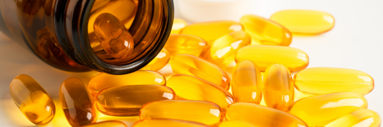
The Omega-3 Lie: Why Fish Oil Capsules Are Failing You — And What Actually Works

The Omega-3 Lie: Why Fish Oil Capsules Are Failing You — And What Actually Works

Here is a question I ask almost every new client: are you taking omega-3 supplements?

The answer is almost always yes. A fish oil capsule, usually. Maybe two a day. They read somewhere it was good for the heart, or their doctor mentioned it, or they picked it up at the pharmacy alongside their multivitamin. They’ve been taking it for months, sometimes years. They assume that because they’re taking it, they’re covered.

Then we run a bloodspot test.

And in the vast majority of cases, the results tell a very different story. Their Omega-3 Index is low. Their omega-6 to omega-3 ratio is badly skewed — not the 2-4:1 that research associates with reduced inflammation and improved health outcomes, but 12:1, 15:1, sometimes higher. Their cell membranes, which should be fluid and responsive, are instead rigid and pro-inflammatory.

The capsule they’ve been taking every morning has not moved the needle.

This is not a fringe observation. It is a systematic problem — one rooted in the quality of most omega-3 products on the market, the form in which they’re delivered, the way they’re stored, and the fundamental fact that without testing, nobody actually knows whether supplementation is achieving anything at all.


Why Omega-3 Status Matters More Than Most People Realize

Before I explain what goes wrong with most supplements, I want to establish why this matters so deeply.

Omega-3 fatty acids — specifically EPA (eicosapentaenoic acid) and DHA (docosahexaenoic acid) — are not simply anti-inflammatory supplements you take to feel better. They are structural components of every cell membrane in your body. EPA and DHA are embedded in the phospholipid bilayer of cell membranes, where they govern fluidity, cellular communication, receptor function, and signaling. This is not a passive role. The composition of your cell membranes determines how efficiently your cells operate at the most fundamental level.

When EPA and DHA are abundant in your membranes, cells are fluid and responsive. Signals pass efficiently. Inflammation resolves appropriately. Neurons fire with precision. The heart maintains stable electrical rhythm. Hormones bind effectively to receptors. When omega-3s are depleted and omega-6 fatty acids dominate instead, the picture reverses: membranes become more rigid and pro-inflammatory, signaling is impaired, and the conditions for chronic disease accumulate quietly over years.

The clinical consequences are well documented. A low Omega-3 Index — the percentage of EPA plus DHA in red blood cell membranes — has been proposed as an independent risk factor for coronary heart disease, particularly sudden cardiac death. An Omega-3 Index in the target range of 8–11% is associated with lower total mortality, fewer major cardiac events, and better organ function — including the brain. Research has consistently linked higher omega-3 membrane content to reduced inflammation, improved mood stability, better cognitive function, healthier pregnancy outcomes, reduced triglycerides, and improved insulin sensitivity.

This is not a supplement story. This is a membrane biology story.


The Ratio Problem Nobody Talks About

There is another dimension of omega-3 status that is even more clinically significant than the Omega-3 Index alone: the omega-6 to omega-3 ratio.

Human beings evolved on a diet where omega-6 and omega-3 fatty acids were consumed in roughly equal proportions — a ratio of approximately 1:1 to 2:1. This balance was maintained through thousands of years of eating wild game, fish, seeds, and plants. Our cellular machinery — the enzymes that convert fatty acids into pro- and anti-inflammatory signaling molecules — was calibrated for this ratio.

The modern Western diet has comprehensively destroyed it. The widespread adoption of vegetable and seed oils — sunflower, corn, soybean, canola — has flooded our food supply with omega-6 linoleic acid. Meanwhile, omega-3 intake has fallen as oily fish consumption has declined. The average Western omega-6 to omega-3 ratio is now estimated at 15:1 to 20:1. Our evolutionary ratio was 1:1.

This matters because omega-6 and omega-3 fatty acids compete directly for the same enzymes. When omega-6 dominates, these enzymes are preferentially used to produce arachidonic acid and pro-inflammatory eicosanoids — the signaling molecules that drive inflammation, immune activation, blood vessel constriction, and platelet aggregation. When omega-3 levels are adequate relative to omega-6, EPA and DHA produce instead the specialized pro-resolving mediators — molecules that actively resolve inflammation rather than simply suppressing it.

The research is unambiguous about the clinical stakes. A ratio of 4:1 in secondary cardiovascular prevention was associated with a 70% decrease in total mortality. A ratio of 2-3:1 suppressed inflammation in patients with rheumatoid arthritis. A ratio of 5:1 had beneficial effects in asthma patients, while 10:1 showed adverse consequences in the same population. The ratio is not just a number — it is a direct reflection of your inflammatory environment at the cellular level.

Most people consuming a modern Western diet, even those taking a standard fish oil capsule, have a ratio far outside this therapeutic range. And most of them have no idea, because nobody has tested it.


Why Most Fish Oil Supplements Don’t Work

This is where I need to be direct about an industry problem that affects most of what is on the pharmacy shelf.

The form matters enormously. Most commercial fish oil supplements are produced in what is called the ethyl ester form. To concentrate EPA and DHA to marketable levels, fish oil is chemically processed — the fatty acids are cleaved from their natural glycerol backbone and reattached to ethanol molecules. This makes the product cheaper to produce and allows higher EPA and DHA percentages per capsule. It also fundamentally alters the molecule.

In the natural triglyceride form — as found in fish — EPA and DHA are attached to a glycerol backbone, exactly as they exist in food and in the body. The digestive system recognizes and processes this structure efficiently. In the ethyl ester form, the body must first cleave the ethanol molecule before it can absorb the fatty acid. This process is significantly less efficient. Research has shown that omega-3s in natural triglyceride form are substantially better absorbed than the ethyl ester form — with one landmark bioavailability study showing that ethyl ester absorption without a high-fat meal can be as low as 20%, compared to far higher rates for triglyceride forms. The ethyl ester processing also releases ethanol during digestion, which may contribute to oxidative stress.

Oxidation is a critical and underappreciated problem. EPA and DHA are polyunsaturated fatty acids — they contain multiple double bonds that are chemically unstable and highly susceptible to oxidation. Once fish oil is processed, exposed to light, heat, or air, the fatty acids begin to oxidize. Rancid fish oil does not just taste unpleasant — oxidized lipids are actively harmful. They generate free radicals, contribute to oxidative stress, and may partially negate the anti-inflammatory benefit the supplement was supposed to provide. Recent analyses suggest that over 60% of omega-3 products on the market exceed recommended oxidation limits at the time of purchase. The fishy burps and aftertaste that many people experience are a reliable warning sign of rancidity.

The dose is almost certainly too low. The standard recommendation of one or two capsules per day — typically delivering 300–500mg of combined EPA and DHA — is rarely sufficient to meaningfully shift membrane composition in someone starting from a depleted baseline with a skewed omega-6:3 ratio. Rebuilding membrane EPA and DHA levels requires sustained, adequate intake over months. At the doses most people take, from a product with compromised bioavailability, very little is actually reaching the membrane in a form the body can use.


Why Testing Changes Everything

This is the argument I make to every client who is taking omega-3 supplements without having ever tested their omega-3 status: you are flying blind.

The Omega-3 Index is to fatty acid status what HbA1c is to blood sugar — a 120-day snapshot of membrane composition that reflects your true long-term fatty acid status, not just what you ate last week. Unlike a serum test that fluctuates with recent intake, the red blood cell membrane test tells you what is actually incorporated into your cellular architecture. It is, in a very real sense, a measure of how your cells are actually built.

The bloodspot testing I use with my clients — a simple finger-prick that can be done at home — measures 11 fatty acids in whole blood and provides six distinct health markers: the Omega-3 Index, the omega-6:3 ratio, protection value, cell membrane fluidity, and mental strength markers. The results arrive within 10–21 days and are delivered securely through an online portal.

What these results reveal is consistently illuminating — and often confronting. I have had clients who have been taking fish oil daily for two years whose Omega-3 Index sits at 3–4%, well below the 8% threshold associated with cardioprotective benefit. I have seen omega-6:3 ratios of 20:1 in people who eat what they consider to be a healthy diet. These are not edge cases — they are the norm in people who have not tested and corrected with a high-quality, bioavailable supplement.

The membrane fluidity marker deserves particular attention. Cell membrane fluidity — the ability of the phospholipid bilayer to flex and allow efficient cellular communication — is a meaningful metabolic marker. Reduced membrane fluidity is linked to impaired glucose transport (a metabolic marker for diabetes risk), reduced red blood cell deformability, increased hemolysis, and oxidative stress. When you understand that the omega-3 composition of the membrane directly governs its fluidity, testing becomes not just useful but clinically essential for anyone managing metabolic health.


What Effective Omega-3 Supplementation Looks Like

When I recommend omega-3 supplementation, I am specific about form, quality, and protocol — because all of these determine whether supplementation actually achieves anything.

Form: Triglyceride form omega-3s are the standard I work with. This is not the cheapest option on the shelf, but it is the option that actually works.

Oxidation protection: High polyphenol content — such as that found in high-quality extra virgin olive oil — provides antioxidant shielding that protects the EPA and DHA from oxidation both in the bottle and in the body. The lipid matrix design of the supplement matters: a MUFA (monounsaturated fatty acid) buffering system, combined with polyphenol antioxidants and reduced oxygen exposure during production, creates a product that retains its integrity through storage and digestion. These are not marketing claims — they are the structural reasons why some formulations produce dramatically better bloodspot results than others.

Dosing: I calculate individual doses based on body weight and baseline bloodspot results. These are personalized, test-driven starting points — not one-capsule-fits-all instructions.

Timing and consistency: Omega-3 supplementation should be taken with a meal containing fat, which optimizes absorption. It should not be taken before exercise, as the acute oxidative stress of training temporarily increases the rate at which EPA and DHA are oxidized in tissues. Consistency over months — not days — is what moves the Omega-3 Index. Most clients see meaningful improvement at the 120-day retest, with target range achievement often requiring 240 days or more from a low baseline.

Testing and retesting: I recommend retesting every 120 days until target range is achieved, and then periodically thereafter. This is not optional. Without retesting, you cannot know whether the intervention is working — and given the variables of individual absorption, existing ratio, dietary omega-6 load, and product quality, the results frequently surprise even those who feel confident they are doing everything right.


The Broader Picture

What the omega-3 bloodspot test reveals is not just an omega-3 level. It is a snapshot of your inflammatory environment — a window into the biochemical conditions that govern whether your immune system resolves inflammation efficiently or sustains it chronically; whether your neurons communicate with precision or struggle through rigid membranes; whether your heart maintains stable electrical rhythm or is more vulnerable to arrhythmia.

The specialized pro-resolving mediators produced from EPA and DHA are not just anti-inflammatory — they actively orchestrate the resolution of inflammation. They signal immune cells to stop producing cytokines, clean up cellular debris, and restore tissue to normal. In a body chronically depleted of EPA and DHA, this resolution phase is impaired. Inflammation is not resolved — it smolders. And chronic, unresolved low-grade inflammation is the biochemical terrain in which virtually every chronic disease takes root.

This is why I consider omega-3 testing foundational — not optional, not a nice-to-have — for anyone I work with who is managing chronic disease, persistent inflammation, metabolic dysfunction, or simply trying to build genuine long-term resilience.

You cannot optimize what you cannot measure. And in almost fifteen years of clinical practice, I have not found a single marker that is more consistently revealing, more consistently modifiable, and more consistently under addressed than omega-3 membrane status.

Take the test. Then act on what you find.


Brigitte Spurgeon works remotely with clients across the US, Canada, Europe, Asia, and Australia. As an Independent Partner with Zinzino, she uses bloodspot fatty acid testing and clinically validated omega-3 supplementation as core tools in her personalized nutrition protocols. To inquire about testing or working together, visit www.brigittespurgeon.com.

This article is for educational purposes and does not constitute medical advice.